
Meniscus Extrusion using Ultrasound
Meniscus extrusion (ME) is defined as the radial displacement of meniscus. This had most commonly been measured using magnetic resonance imaging (MRI) and had been prospectively correlated with development of tibiofemoral cartilage loss, premature osteoarthritis, and degenerative subchondral marrow changes.
Musculoskeletal ultrasound is readily available in clinics and is a cost-effective imaging modality that is increasingly utilized in evaluation of musculoskeletal injuries. Major advantage of ultrasound compared to MRI is the ability to dynamically assess the meniscus extrusion under stress. This allows for real-time assessment of ME under varying loading conditions of the knee joint, as opposed to clino-static evaluation typical of MRI. Musculoskeletal ultrasound had been utilized with early success in assessing structural information of the lateral and medial meniscus. Current work in the lab is focused on correlating meniscus extrusion to various meniscus injuries.
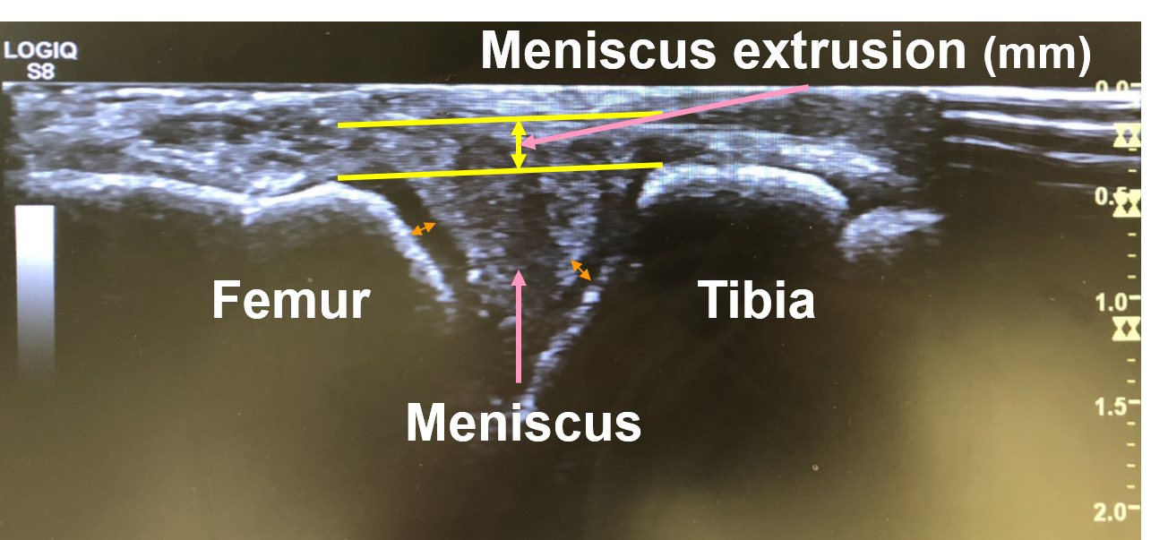
Image 1: Ultrasound image displaying extrusion of the medial meniscus

Image 2: Diagram showing medial meniscus extrusion