Biomechanical Factors Influencing the Successful Non-operative Treatment of Rotator Cuff Tears
Rotator cuff tears are a significant clinical problem that affect greater than 1/3 of all people over the age of 60 and can cause severe shoulder pain and loss of function. While physical therapy is commonly prescribed for the initial treatment of many rotator cuff tears, it has been found to be ineffective in as many as 25 to 50% of all cases, requiring surgical intervention. Since there is no "one size fits all" approach to treating rotator cuff tears, which can come in many different sizes, shapes, locations, and chronicities, it is necessary to better understand the biomechanical reasons behind the failure of physical therapy in order to reduce treatment time and costs. Therefore, the objective of this study is to use in vivo, cadaveric, and computational approaches to investigate the effects of physical therapy and initial tear characteristics on tear propagation and alteration of glenohumeral kinematics.
POETT Study
Under the direction of Dr. Richard Debski, Dr. James Irrgang, and Dr. Volker Mushal, a five-year study called, “Predicting the Outcome of Exercise Therapy for Treatment of Rotator Cuff Tears,” or POETT, aims to develop diagnostic methods to allow surgeons to determine whether physical therapy or surgery is the most effective initial treatment of rotator cuff tears. Through a collaboration with the Department of Physical Therapy and co-investigator Dr. William Anderst's Orthopaedic Biodynamics Laboratory in the Department of Orthopaedic Surgery, we are working to collect in vivo glenohumeral joint kinematic data from 100 patients with rotator cuff tear before and after they have gone through a 12-week program of physical therapy. Bi-planar x-ray images provide quantitative measurements of shoulder motion during activities of daily living and before and after physical therapy. This information will be combined with isometric strength data, shoulder function questionnaires, and ultrasound measures of tendon thickness and tear size taken over time during physical therapy sessions to improve our understanding of the effects of physical therapy for rotator cuff tear treatment on glenohumeral joint health. The long-term goal of this $2.79 million NIH-funded study is to perform a clinical trial to determine whether predictions determining whether patients should initially undergo non-operative or surgical treatment at time of diagnosis make a difference in treatment outcomes.

Related Publications:
Miller RM, Popchak A, Vyas D, Tashman S, Irrgang JJ, Musahl V, Debski RE. Effect of Exercise Therapy for the Treatment of Symptomatic Full-Thickness Supraspinatus Tears on in vivo Glenohumeral Kinematics. J Shoulder Elbow Surg. 2016 Apr; 25(4):641-9.
http://www.ncbi.nlm.nih.gov/pubmed/26620280
Cadaveric Testing
Extensive biomechanical testing on cadaveric human rotator cuff tendons under cyclic loading conditions is being performed in order to assess the effects of tear location and chronicity on tear propagation. Comparisons of tear location between the anterior and central supraspinatus tendon have thus far shown no significant difference in load required to reach 2 cm of propagation, though anterior tears have been found to propagate earlier than central tears, demonstrating an effect of tear location on tear propagation.
Strain measurements made using a video tracking system have helped to elucidate tear propagation based on location and direction of maximum principal strains on the tendon surface. Ongoing work on cadaveric tissue with pre-existing chronic tears is being performed to compare with surgically created "acute" tears to assess the effects of tear chronicity on tear propagation. Additionally, histological analysis has been performed to investigate the localized degeneration in rotator cuff tendons, and has found increased amounts of lipoid infiltration in the tendon near the myotendinous junction.


Related Publications:
Araki D, Miller RM, Fujimaki Y, Hoshino Y, Musahl V, Debski RE. Effect of Tear Location on Propagation of Isolated Supraspinatus Tendon Tears During Increasing Levels of Cyclic Loading. J. Bone Joint Surg. Am. 2015 Feb 18; 97(4):273-278.
http://www.ncbi.nlm.nih.gov/pubmed/25695976
Miller RM, Fujimaki Y, Daisuke A, Musahl V, Debski RE. Strain Distribution due to Propagation of Tears in the Anterior Supraspinatus Tendon. J. Orthop. Res. 2014 Oct;32(10):1283-9.
http://www.ncbi.nlm.nih.gov/pubmed/24985532
Computational Modeling
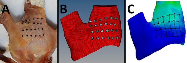
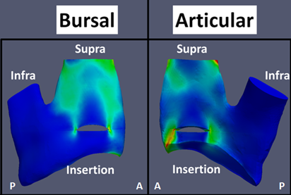
Ongoing work with Dr. Maiti in the Department of Bioengineering will result in the development of a finite element model to simulate tear propagation in rotator cuff tendons for a variety of tear characteristics. Cohesive elements have been used with subject-specific tendon geometries and material properties to model propagation of tears that vary in size, shape, and location while varying material properties to approximate differences due to tissue degeneration. Biomechanical testing combined with high-resolution strain maps of the bursal and articular surfaces of tendon have allowed for validation of the model surface strains. Modeling has shown similar results of experiments, indicating that tears in the anterior third of the tendon and tears greater than 1 cm in size are most at risk for propagation.
Related Publications:
Thunes J, Miller RM, Pal S, Damle S, Debski RE, Maiti S. The Effect of Size and Location of Tears in the Supraspinatus Tendon on Potential Tear Propagation. J Biomech Eng. 2015 Aug 1;137(8). doi: 10.1115/1.4030745. Epub 2015 Jun 23.