Ankle Joint Biomechanical Testing
Ankle Syndesmosis
High ankle sprains are common in contact sports such as football and soccer. A high ankle sprain typically occurs when an external rotation force is applied to the ankle in prone position. This type of sprain is characterized by damage to the interosseous membrane and syndesmotic ligaments that bind the tibia and fibula. The primary goal of this project is to characterize fibular and talar motion after syndesmotic injury and surgical fixation with simulated weight-bearing. Surgical fixation methods tested in this project include: tricortical screw constructs, suture button constructs, and hybrid constructs.
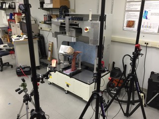
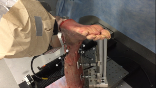
Testing of a full lower leg cadaveric specimen (shank and foot) introduces additional constraints not present in knee joint testing. First, three rigid bodies (tibia, fibula, and talus) must be tracked to determine kinematics of the high ankle joint. Second, a larger specimen must be fastened to the robot for testing, which required the construction of custom clamps to rigidly mount the specimen to the robot. In order to conduct biomechanical testing of the high ankle joint, an external, 3-dimensional motion tracking system (OptiTrack) is being used to record the motion of the full length fibula.
*Photo contains sensitive content which some people may find offensive or disturbing. Click to reveal.

