
Picture Perfect
")
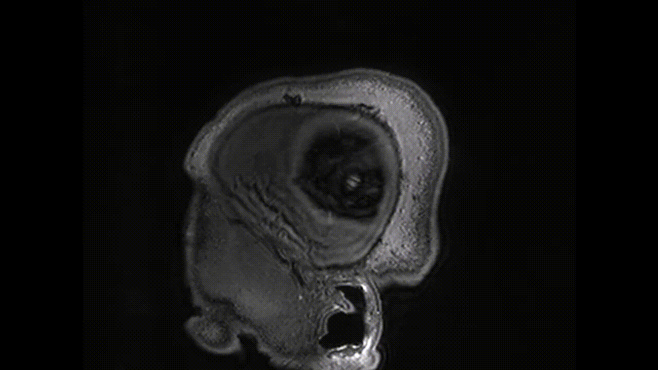
Jonathan Vande Geest distinctly remembers the exact moment he knew he needed a two-photon microscope. A colleague had pulled up a surprising image on his screen: a dense, luminous web of collagen fibers, rendered in detailed 3D. Vande Geest asked: how long did it take to prepare the sample? How many hours of fixing, freezing, sectioning, and staining did it take?
“None,” his colleague said. “That's just a piece of tissue I put under the microscope.”
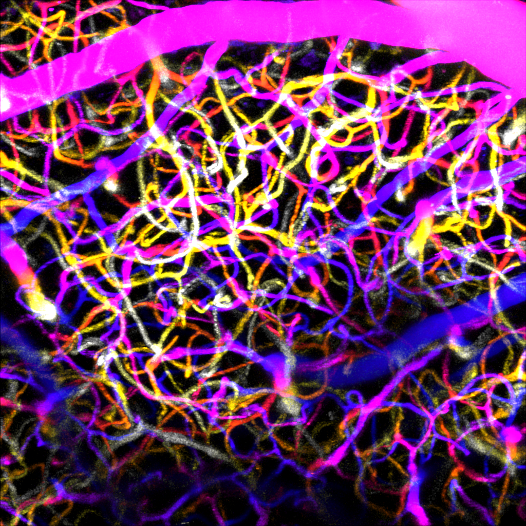
"My jaw dropped," said Vande Geest, professor of bioengineering at the University of Pittsburgh’s Swanson School of Engineering. “The ability to image collagen in 3D without fixing it first means I can look at it under mechanical load, and I can also watch it grow and change. As a soft tissue biomechanist, there is really no better thing."
Two-photon microscopy is just one of the many impactful imaging modalities that are put to work across Pitt’s campus. Bioengineering researchers like Vande Geest have built the tools and expertise to ask and answer questions that couldn't have been posed before, from the tiniest cellular movements to the complex architecture of the aging human brain.
Two-Photon Microscopy
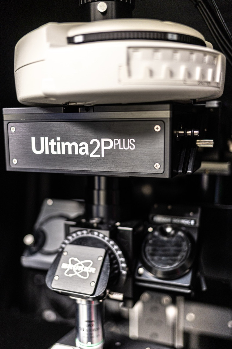
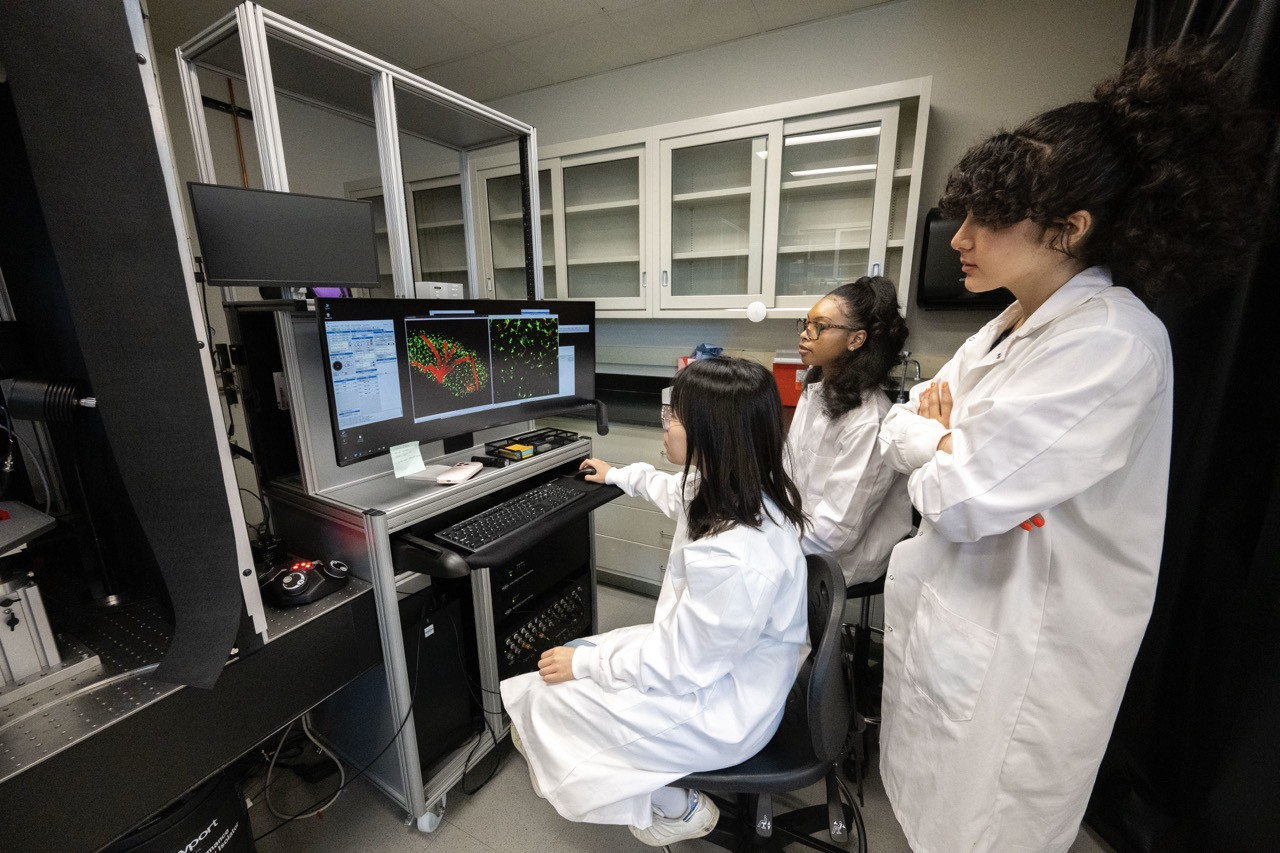
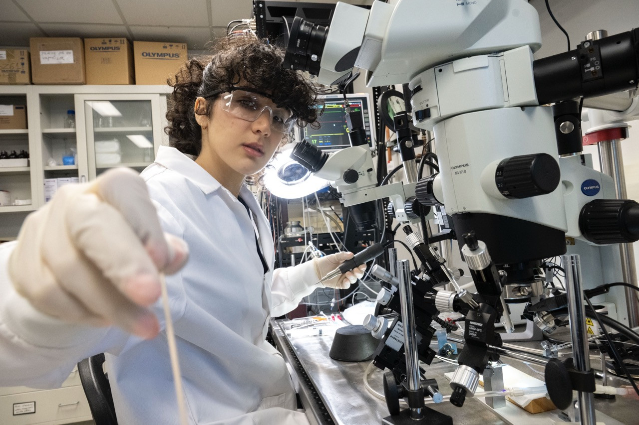
Two-photon microscopy fires pulses of infrared light into tissue. Unlike conventional light, infrared travels deep without scattering, but it only produces fluorescence at the precise focal point where the beam converges. At Pitt’s Center for Biotechnology and Bioengineering, you willl find a few of these microscopes, each customized for a different scientific purpose. One of these devices is helping Takashi (TK) Kozai, Ernest E. Roth professor of bioengineering, peer into the brain to analyze a largely unexplored corner of neuroscience.
"Brain tissue is like a very foggy, hazy environment, and neurons aren't on the surface level, so we have to image a little deeper to actually see them," said Kozai. “That’s where two-photon comes into play and can help us see very specific areas of the brain.”
Kozai’s team studies what happens when devices like electrodes are implanted, not just to neurons, but to the surrounding community of glial cells. Because glial cells don't generate electrical signals that electrodes can detect, they are effectively invisible to conventional methods.
"But with 2P, you can label different subtypes of neurons or other cell-types so that you can see which ones are activated under which type of stimulus," Kozai said. "You can decode beyond what an electrode can decode."
Kozai’s microscope is available for researchers of all disciplines to use, which helps him better understand how to improve the long-term performance of implanted devices toward the ultimate goal of restoring motor function in people with spinal cord injuries or treating vision loss in patients who are blind. Vande Geest, on the other hand, uses his scope for different terrain entirely: studying the extracellular matrix of soft tissues found in the eye, blood vessels, peripheral nerves, and more. Collagen, the structural protein that gives these tissues their mechanical character, produces a signal under two-photon illumination that makes it visible without any dye or label. More importantly, it stays visible while the tissue is alive, under load, and changing over time.
"I study how tissues are built and how they change, and this is the system that lets me do it. I can image it, deform it, and image it again," he said. “I can literally watch collagen remodel.”
Vande Geest has also reconfigured his microscope to function as a 3D printer that can fabricate structures at the scale of individual cells. The same instrument that shows the architecture of a tissue sample can now print scaffolds that replicate it, opening a path toward implantable tissues that could treat vascular and ocular disease.
“A majority of the intellectual property (IP) I've developed involves benchtop platforms for mimicking human disease in 3D tissue culture," Vande Geest said. "We can take human stem cells, differentiate them, and assemble them into something that might actually tell a clinician whether a patient should be treated more aggressively for something like glaucoma, and that specialized platform gives you information that clinical measurement alone just can't."
A Picture’s Worth
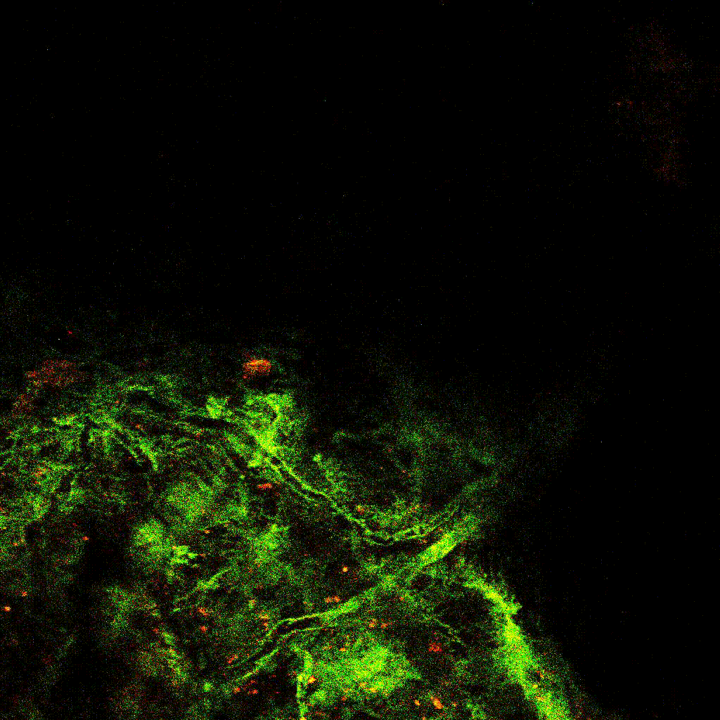
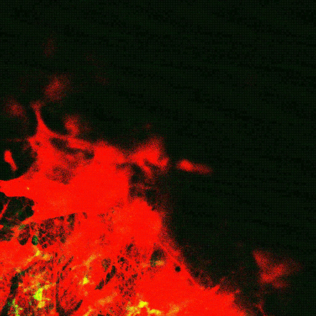
For Lance Davidson, William Kepler Whiteford Professor of bioengineering, collecting an image is only the beginning. Using traditional confocal microscopy, which uses focused laser light to build three-dimensional images one optical slice at a time, his lab studies how cells rearrange inside tissues under mechanical stress, using frog embryos as a model system. These embryos are optically transparent and mechanically tractable in ways that make them ideal for stretching, compressing, and perturbing living tissue while imaging what happens inside it.
"We use a lot of microscopy to collect images, and it's all light microscopy, not anything too complex." Davidson said. "But we combine these tools with molecular genetic approaches, so we can install a fluorescent protein within a cell or tissue that indicates where forces are generated, or how material properties adapt to the environment.”
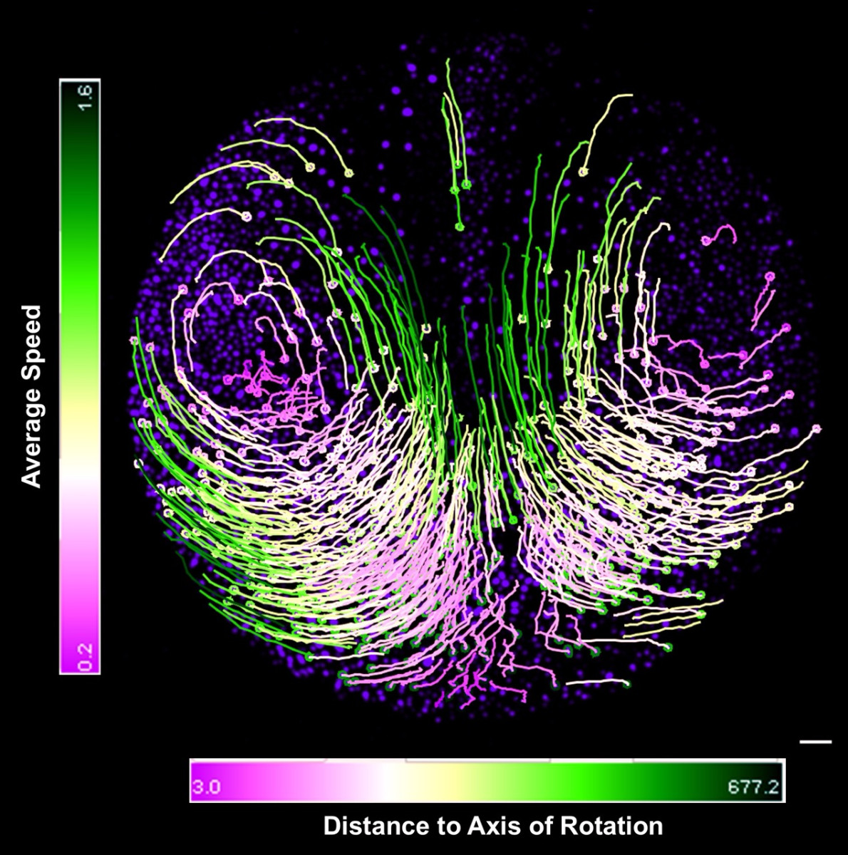
After collecting time-lapse sequences of cells moving through a tissue, Davidson’s team builds custom image processing pipelines that segment individual cells, track which cells are neighbors at each time point, and quantify how frequently and in what direction those relationships change. In a recent study, that analysis revealed something novel in the raw footage: a pair of counter-rotating flows inside a developing tissue, moving in opposite directions.
"Once we processed it, we could see these really incredible flows.” Davidson said. “They looked like tropical cyclones or vortices on the surface of the sun, and so we turned to quantify the strength of those rotations using tools borrowed from astrophysics."
For Davidson, however, the most important work happens after the microscope turns off. A striking image is still just a picture until it's been broken down, processed, and reduced to something a statistician can work with.
"The adage ‘a picture is worth a thousand words’ is actually quite terrible for science," Davidson said. “Numbers are like currency, and we use images to get that currency, so you really want each picture to be worth a single number."
Sound as Sight
Of all the imaging modalities in use, ultrasound may be the easiest to underestimate. It's one of the oldest, the most affordable, the most common, and it doesn't carry the glamour of a two-photon beam. But across the university, researchers are finding new uses for it that go far beyond the routine.
Kozai's lab, for instance, recently found that low-intensity ultrasound can reduce the glial scarring that builds up around implanted brain electrodes, keeping signals clearer over time and opening new possibilities for modulating non-neuronal brain cells. In addition, Kang Kim, professor of bioengineering and medicine at UPMC’s Heart and Vascular Institute, is also pushing ultrasound far beyond its typical uses.
"Ultrasound has been out there for decades," Kim said. "It's safe, non-invasive, and real-time. But because of how well we understand physics, we can now start to combine it with other modalities and push it in directions people didn't think were possible."
One of Kim’s current projects involves a catheter-based imaging system designed to peer inside blood vessels at the microscopic level. The project addresses plaque vulnerability, assessing which arterial plaques are likely to rupture and send a clot toward the heart or brain. One signature of a dangerous plaque is the presence of tiny microvessels growing within it, and to detect them, Kim's team developed an intravascular probe and signal processing approach that can image those structures at scales previously considered beyond the physical limits of the modality.
"We claim this is one of the first kinds of intravascular super-resolution imaging of microvessels," Kim said. "There are emerging fields even beyond imaging where ultrasound can be used. It is not just a tool for looking; it is becoming a tool for doing."
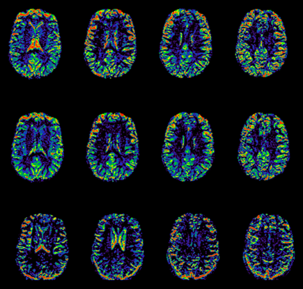
MRI and the Whole-Brain Picture
While microscopes allow for cellular or tissue analysis and ultrasound can peer inside vessels and soft tissue in real time, there's a hard physical limit to how deep light can travel. When researchers need to see the structure, connectivity, and metabolic activity of the entire brain, they turn to MRI.
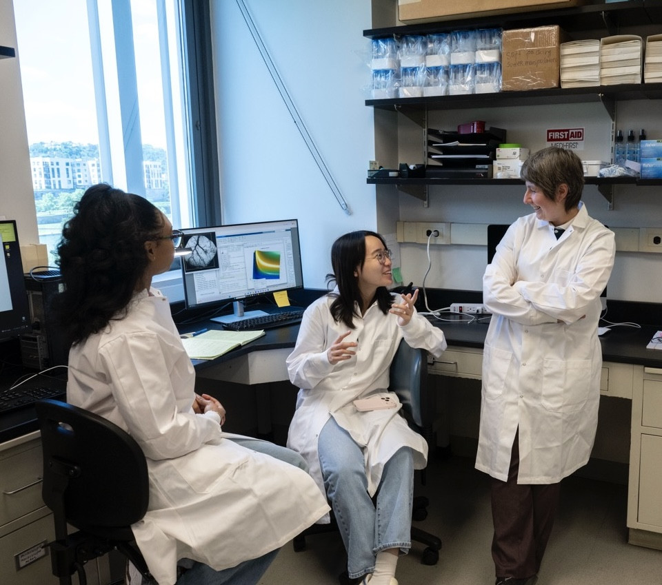
Bistra Iordanova uses a combination of optical imaging techniques in her work, but uses structural and functional MRI (fMRI) in tandem with optical approaches to get a window into brain-wide activity that no light-based system can match.
"With optics, we can get the cell resolution, but MRI covers the entire brain at once, which makes it indispensable for questions about large-scale connectivity and system-wide disease," said Iordanova, assistant professor of bioengineering. “Structural MRI shows the size and shape of brain regions and how they change with age, while fMRI tracks blood flow oxygenation as a proxy for neural activity in real time.”
Using these techniques, she's currently working on a methodologically unusual project: directly comparing data between mice and humans to help design multiscale models of how brain metabolism can change the risk for dementia. This approach spans three scales: two-photon microscopy to quantify blood cell velocity, neural activity, and metabolite levels at the cellular level; wide-field imaging to capture how mitochondrial activity moves across cortical networks; and whole-brain MRI to explore how energy metabolism shapes functional connectivity across both animal models and human cohorts.
"To make these comparisons work, we transform the imaging data to the same parameters," Iordanova said. "In the human brain, a vessel might be two centimeters long, but in a mouse brain, it’s two millimeters. And a mouse only lives two years while a human lives 80. This kind of cross-species translation can actually be quite difficult, but it's where the real clinical relevance lies.”
Building the Brain Scan
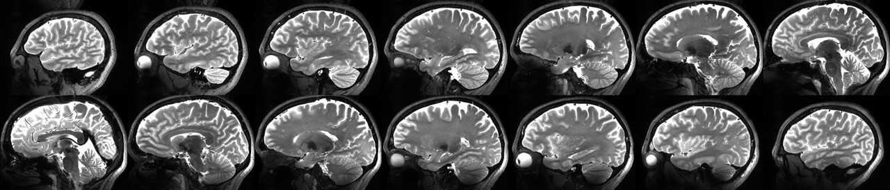
While many researchers like Iordanova are using the existing MRI scanners in their work, Tamer Ibrahim, professor of bioengineering, has spent more than 20 years engineering the technology itself.
For MRI, the stronger the magnetic field, the greater the signal-to-noise ratio, and the finer the structural detail that becomes visible. But scanners at a high magnetic field like 7 Tesla come with a serious engineering problem: the interactions between high-frequency electromagnetic waves and human tissue can create dead zones in the image, or regions of the brain that simply produce no signal.
Ibrahim's lab, the 7 Tesla Bioengineering Research Program (7TBRP), has solved this problem with a custom radiofrequency coil system called Tic-Tac-Toe, and its second-generation successor, the Tac G2, introduced in 2022. The Tac G2 is, by Ibrahim's account, the only system in the world that has comprehensively eliminated the signal void problem, allowing researchers to run any type of MRI study at 7T without imaging barriers.
"There are significant challenges when scanning at 7T," Ibrahim said. "But our anti-claustrophobia Tac G2 coil system is, to my knowledge, the only one in the world that has successfully and comprehensively solved this problem.”
The practical consequences are substantial. A study published in Human Brain Mapping from Ibrahim's team, comparing 3T and 7T performance across 350 healthy adults, found that 7T produces stronger correlations with age-related brain changes across every measure examined such as cortical volume, subcortical volume, white matter, cortical thickness. More importantly, it found a study that would require 350 participants at 3T could achieve the same statistical significance with approximately 100 participants at 7T.
That efficiency gain means that studies that were previously too expensive, too slow, or too logistically demanding to conduct become feasible. Since the Tac G2's introduction, it has been used in more than 2,500 human scans, already surpassing its predecessor's total in less than half the time. More than 40 NIH-funded studies across aging, psychiatry, neurology, and neuroscience are currently running on the system.
"When our coils are used in human studies, it's incredibly rewarding, far more rewarding than just publishing a paper," he said. "We're developing devices that clinicians and scientists use, and the result isn't just pretty pictures. We're not making something that just could be used some time in the future, we’re impacting human life now."
The Full Picture
These researchers work with different tools, different tissues, and different diseases, yet the opportunity for collaboration between modalities seems to increase by the day. A shared conviction ties all of their work together: there is no perfect imaging modality. Every technique involves tradeoffs between resolution and depth, speed and sensitivity, invasiveness and detail. For Iordanova, those limitations are precisely what makes the field so interesting and allows for such innovation.
"If you don't have solid image analysis, it doesn't matter if you have a fancy machine," she said. "That's the beauty of bioengineering - you get to reach into any pocket you want. Optics, electrical engineering, image processing, artificial intelligence. The biology department says stick to cells, the electrical engineering department says just do the signal processing, but bioengineering lets you have it all."
Interested in using a 2P microscope for your research project? Contact TK Kozai for more information at tdk18@pitt.edu.