Project 4:
Stimulation with Neural Electrodes
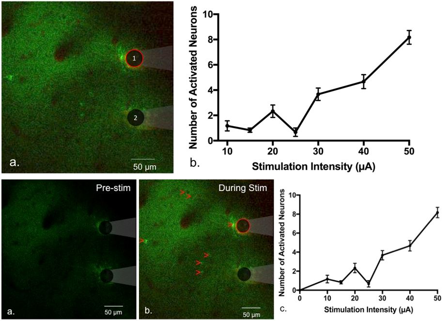
Stimulation through microelectrodes offers opportunities to gain high spatial selectivity, but imposes unique challenges to the electrode materials. In order to achieve effective activation of the neural tissue, sufficient charges need to be injected from microelectrodes without exceeding the potentials at which irreversible electrochemical reaction occur. While the safety of stimulation has been extensively studied for metal electrodes, the stimulation parameters for emerging materials (e.g. conducting polymers with various dopants) still need to be optimized to achieve effective stimulation at with minimal risk. With the use of imaging tools such as the two photon microscopy and meso-scale florescent microscopy and transgenic animals such as GCaMP mice we can observe and quantify the effect of electrical stimulation on neural elements.

Figure 1. Two-photon-imaging of neurons before (a) and after (b) stimulation via a PEDOT/CNT electrode (red outline). GCaMP is expressed in neurons, shown in green. Blood vessels are labeled by sulforhodamine 101 (SR101) in red. Red arrows point to electrically evoked neurons (c) The number of electrically evoked neuronal cell bodies increases as a function of stimulation current amplitude using the pulse shape (n = 6 trials, error bar = SEM).